Signs and symptoms
- Correct diagnosis and fast
- Irreversible necrosis within hours
- Signs of occluded artery in the face
- Particularly painful injection
- Spasm of the artery and area of pallor in the skin
- Sensation of pressure after injection
- Capillary refill should be done as standard after every procedure, negating the risk of sending the patient home and something present later on
- If unsure, prick area of skin with needle, should experience blood, if not be concerned
- Patient returns with complaint 6-24 hours later if sent home with no concern
- Significant pain and sometimes bruising
- Dusky, grey mottled skin, patient must be examined in person
- Cap refill slow or non-existent
- 24 hours pain will increase with inflammation to dying tissues
- 48-72 hours signs of full-blown necrosis will ensue
- Dark/black tissue appearance with small pustules also arising
The following 5 steps can aid in the diagnosis quickly
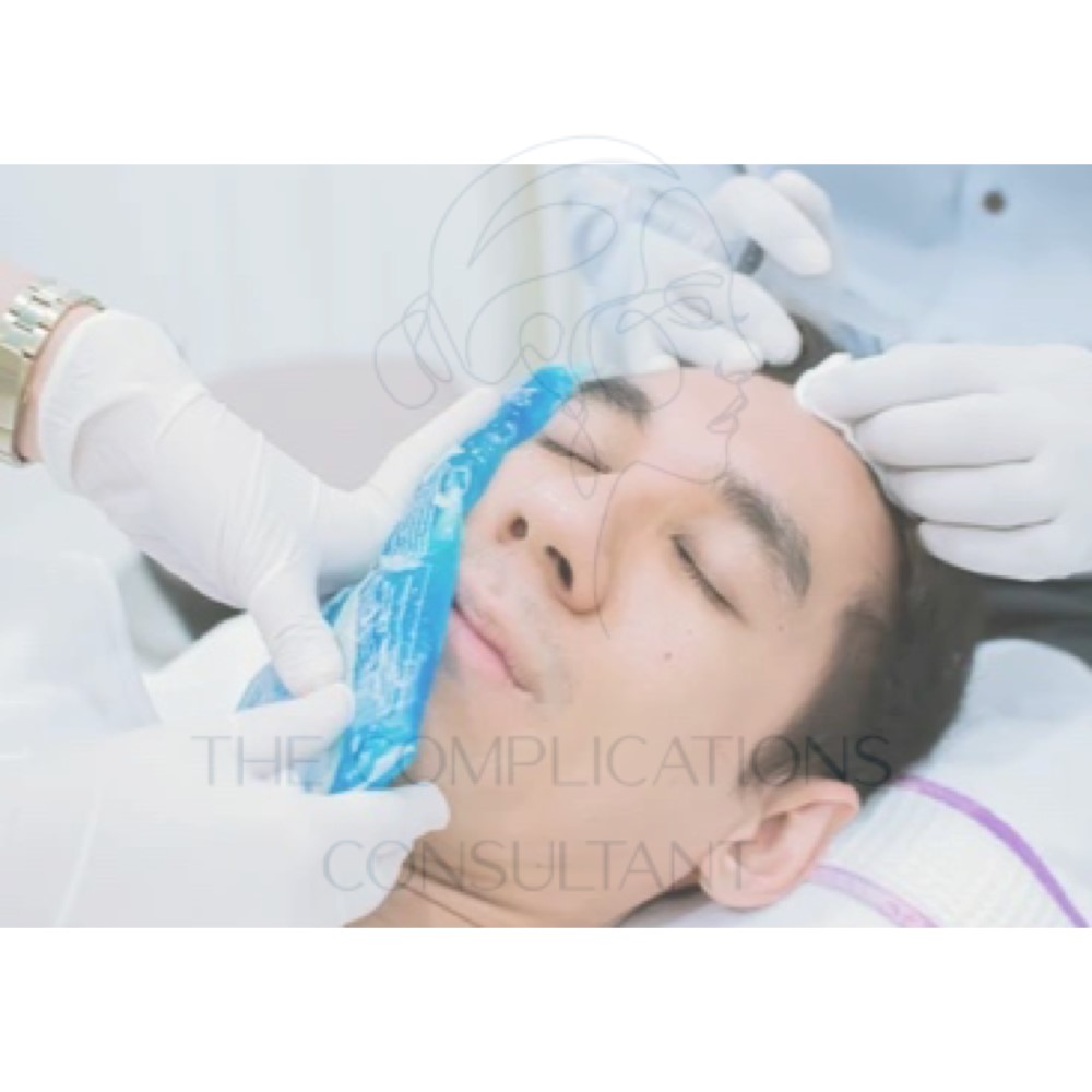
1. Capillary refill is key! The use of clinical photographs when seeking help and diagnosis is of course an option. However accurately documenting the capillary refill time will be paramount in correctly diagnosing a vascular occlusion. Less than two seconds is an acceptable refill time, more than this then the tissues and cells may be jeopardised.
2. Use an untreated part of the body as a control or standard to measure the CR up against. Tis will evidence what is in the normal range for your patient as an individual. Do this at the same time so it’s easy to compare.
3. Compress with firm pressure for at least 5 seconds. Always compress the area firmly for at least 5 seconds otherwise not enough blood is squeezed away from the area.
4. If you decided you have in fact caused a VO then the best way forward is to map out the pathway of the artery with a pencil onto the patients face using your anatomical knowledge. Whatever artery it may involve follow the pathway rigorously and consider where it could be blocked, include harder to see areas like the columella of the nose. The use of a cotton bud for capillary refill checks in these areas will also be helpful as they are smaller than your finger and can get into harder to reach places.
5. The pin prick test should be adopted if the patient has harder to see areas of the face as a result of pigmentation or freckles, this will give reassurance hopefully that blood flow is present.
Additional presentation to note will be puffiness of the area not being drained, discolouration with the build-up of blood in the tissues.