Causative types of injury
Let’s look in detail at the types of injury that may cause visual loss.
OPTHALMIC ARTERY OCCULSION
- Most severe
- Doesn’t affect central nervous system as vessel supplies other vessels in the orbit
- As a result, can affect muscles of the eye also
- Effects entire blood supply of the globe and all parts of the retina
GENERALISED POSTERIOR CILIARY ARTERY
- Occlusion with relative central retinal artery is not massively prominent
MACULA
- Only part of retina not supplied by central retinal artery
- Supplied by posterior ciliary vessels
- For this reason, occlusion effects macula
- Loss of central and acute aspects of vision
- Will be less of an issue on peripheral vision
CENTRAL RETINAL ARTERY OCCLUSION
- Affects blood supply of entire retina
- Small Macula portion supplied by posterior ciliary artery
BRANCH RETINOL ARTERY OCCULUSION
- Damage causes partial peripheral loss of vision
- Caused by emboli effecting central retinol artery
ARTERIAL PATHOLOGY
ANTERIOR ISCHEMIC OPTIC NEUROPATHY
- Cause by damage and loss of bloody supply to the anterior optic nerve
- Occurs at the area of the optic disc
- Variable degrees of vision loss experienced
- Above depends on amount and extent of trauma
POSTERIOR ISCHEMIC OPTIC NEUROPATHY
- Caused by damage to the blood supply of the posterior portion of the optic nerve.
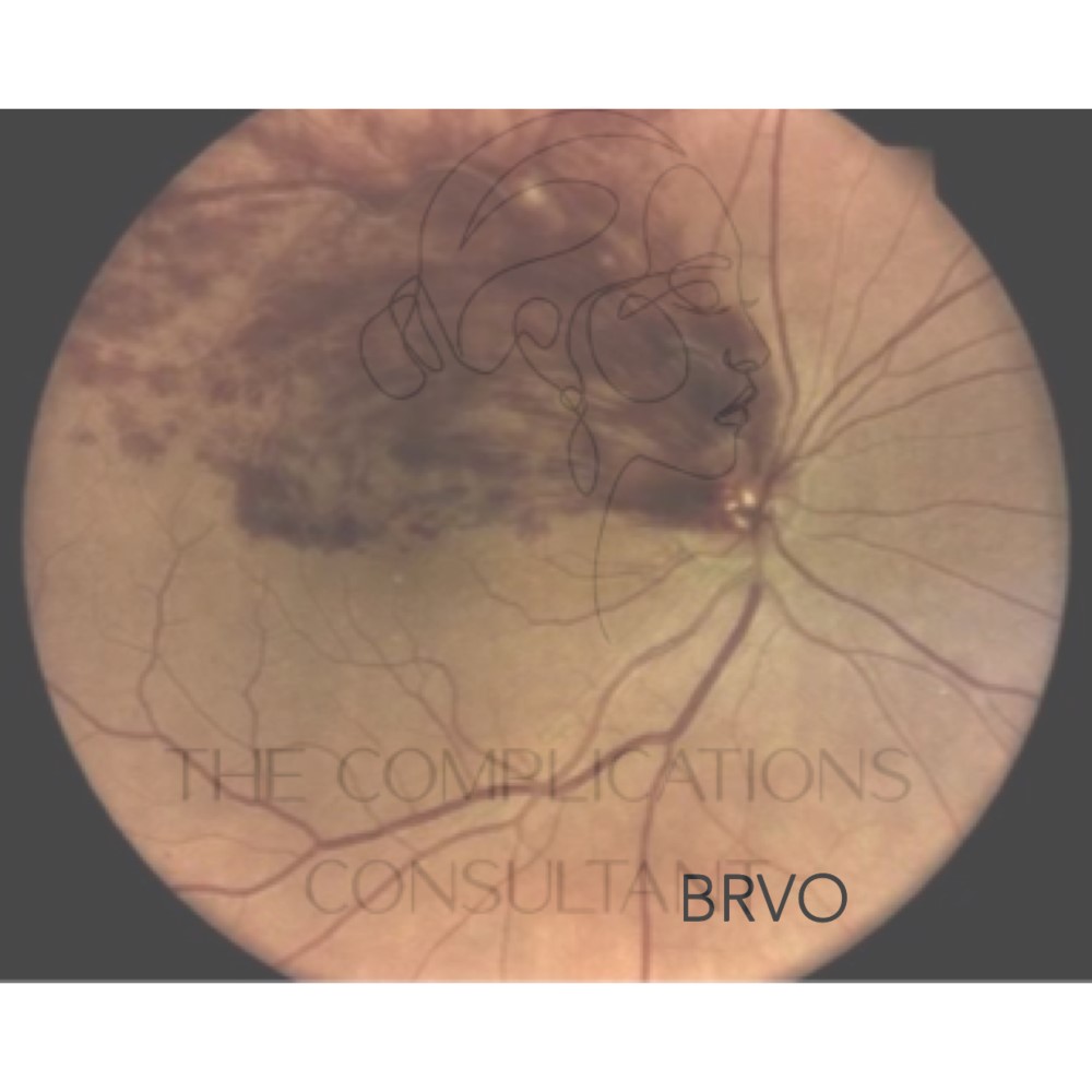
VENOUS PATHOLOGY
There are four main vessels that drain the posterior quadrants of the eye, if these become blocked or compressed and drainage is prohibited, the retina can be damaged.
This can affect one or more these four vessels and is known as BRANCH RETINAL VEIN OCCLUSIONS. The central retinal vein can also be affected by this, which can predispose the cavernous sinus to occlude. As seen previously in the flow diagram, this anatomical structure is responsible for retinal blood drainage. This is a high-risk area of vision loss in relation to blockage.
The temporal vein is a high-risk area for vision loss if damaged, compressed or blocked by dermal filler. Reported incidence of such is a good reminder to be very careful and mindful of anatomy when conducting treatments in this area.
The above mentioned types of occlusion and the anatomy they affect will translate into different types of classification. These directly help identify the clinical presentation and scenario surrounding the incidence and are as follows:
TYPE I
A shorthand for blindness injury that occurs in isolation
TYPE II
Blindness with corresponding damage to the levator muscles of the eyelid
TYPE III
Blindness with injury to the ocular muscles causing ophthalmoplegia
TYPE IV
Ophthalmoplegia with ptosis of both eyelids